How Our Eyes Work and What They’re Made Of:
The human eye is often described as one of the most remarkable organs in the human body. It allows us to experience the world in vibrant color, depth, and motion. But how does this incredible organ actually work? In this article, we’ll dive into the fascinating make and structure of the human eye, also exploring how each part contributes to our sense of sight.
A Quick Overview of the Human Eye
Imagine the eye as a highly sophisticated camera, constantly capturing images and sending them to the brain for processing. The eye’s structure is complex, comprising several key parts that work in harmony to allow us to see clearly. Each component plays a crucial role, from focusing light to converting it into signals that the brain understands. Here’s a closer look at how the eye is built.
The Outer Structure of the Eye
The eye is enclosed by several protective layers and components that ensure it remains functional and safe.
1. Cornea
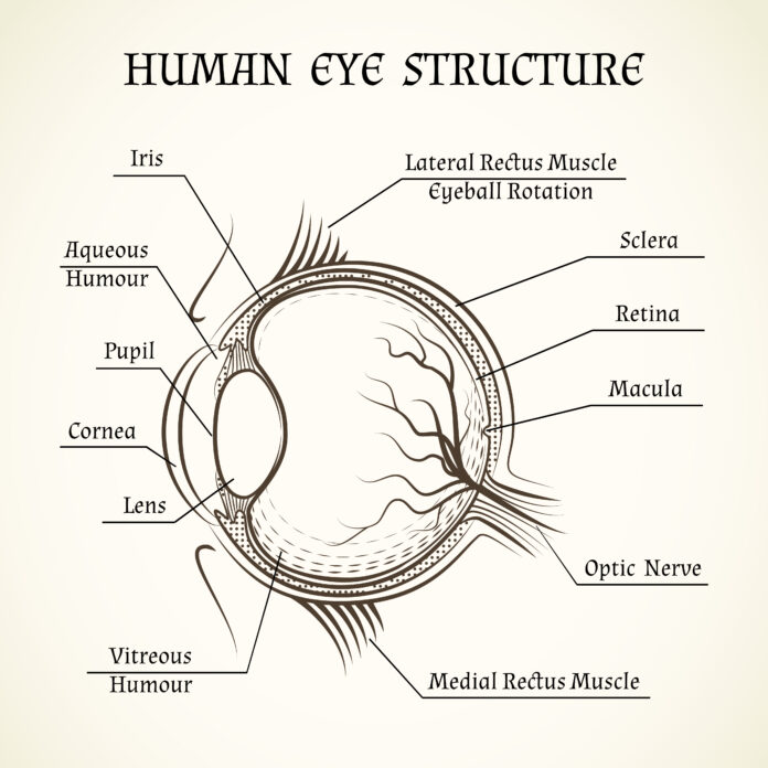
The cornea is the transparent, dome-shaped surface that covers the front of the eye. It’s the first point of contact for light entering the eye and plays a critical role in focusing that light onto the retina. The cornea is composed of five layers, each with a specific function. It’s both strong and delicate, providing a protective barrier against dust, germs, and other potential hazards. Think of the cornea as the eye’s window, allowing light to enter while keeping unwanted elements out.
2. Sclera
The sclera is the white, opaque part of the eye that surrounds the cornea. It’s made of tough, fibrous tissue that protects the eye and also gives it its shape. The sclera is like the eye’s outer shell, providing structural support and anchoring the muscles that control eye movement.
3. Conjunctiva
The conjunctiva is a thin, transparent membrane that covers the sclera and the inner surface of the eyelids. It helps keep the eye moist by producing mucus and tears and also serves as a protective layer against infections.
The Middle Layer: Choroid, Ciliary Body, and Iris
Beneath the outer layer of the eye lies the middle layer, which is rich in blood vessels and crucial for the eye’s nourishment.
1. Choroid
The choroid is a layer of blood vessels located between the sclera and the retina. It supplies oxygen and nutrients to the eye, particularly the retina, which is also essential for maintaining healthy vision. Although the choroid also helps absorb stray light, preventing it from scattering inside the eye, which would otherwise blur our vision.
2. Ciliary Body
Connected to the choroid, the ciliary body is a ring-shaped structure that produces aqueous humor (a clear fluid that fills the front part of the eye). It also contains the ciliary muscle, which controls the shape of the lens, allowing us to focus on objects at varying distances—a process known as accommodation.
3. Iris
The iris is the colorful part of the eye, often seen in shades of blue, green, brown, or hazel. It functions like a camera’s aperture, controlling the size of the pupil to regulate the amount of light entering the eye. In bright light, the iris contracts, making the pupil smaller to reduce light entry. In dim conditions, it expands the pupil, allowing more light in. This automatic adjustment helps protect the retina from excessive light and improves vision in low-light conditions.
The Inner Workings of the Eye
Now, let’s look at the inner parts of the eye where the magic of vision truly happens.
1. Lens
Situated just behind the iris, the lens is a transparent, flexible structure that also focuses light onto the retina. It works in tandem with the cornea to fine-tune the focus, enabling us to see objects clearly at different distances. The lens changes shape through the action of the ciliary muscle: it flattens to focus on distant objects and becomes rounder to focus on closer ones.
2. Retina
The retina is perhaps the most critical part of the eye—a thin layer of tissue lining the back of the eye that functions like the film in a camera. It contains millions of light-sensitive cells known as photoreceptors, which come in two main types: rods and cones.
- Rods are responsible for vision in low light conditions. They’re highly sensitive to light but do not perceive color, making them crucial for night vision.
- Cones are responsible for color vision and work best in bright light. There are three types of cones, each sensitive to different wavelengths of light corresponding to red, green, and blue. Together, they allow us to see the full spectrum of colors.
The retina captures light and converts it into electrical signals, which are then sent to the brain via the optic nerve.
3. Macula and Fovea
The macula is a small, central area of the retina responsible for sharp, detailed vision. At the very center of the macula is the fovea, a tiny pit packed with cones that provides the clearest vision and is essential for tasks like reading and recognizing faces.
4. Optic Nerve
The optic nerve is the crucial connection between the eye and the brain. It transmits visual information from the retina to the brain, where it is processed and interpreted as images. This neural highway is like a high-speed internet connection, relaying vast amounts of data in fractions of a second.
The Fluid Systems: Aqueous and Vitreous Humors
Two types of fluid help maintain the eye’s shape and internal pressure while providing nutrients to various eye structures.
1. Aqueous Humor
The aqueous humor is a clear, watery fluid that fills the space between the cornea and the lens. It helps maintain the eye’s shape, provides nutrients to the cornea and lens, and removes waste products. The continuous production and drainage of aqueous humor are vital for maintaining proper intraocular pressure, which is necessary for the eye’s overall health.
2. Vitreous Humor
Behind the lens lies the vitreous humor, a gel-like substance that fills the large cavity of the eye. It helps maintain the eye’s shape and also keeps the retina in place by exerting slight pressure against it. The vitreous humor is composed mainly of water but also contains proteins and collagen fibers, giving it a jelly-like consistency.
The Protective Mechanisms of the Eye
Our eyes are equipped with several natural defenses to protect them from harm and maintain their functionality.
1. Eyelids and Eyelashes
Infact eyelids act as protective shields, closing reflexively to guard against bright light, foreign particles, and potential injuries. Eyelashes help filter out dust and debris, while the blinking action spreads tears across the surface of the eye, keeping it moist and free from irritants.
2. Tear Film
The eye’s surface is coated with a thin layer of tears, known as the tear film. This film is essential for keeping the eye moist, providing nutrients, and creating a smooth surface for light to enter. It consists of three layers: an oily layer that prevents evaporation, a watery layer that hydrates, and a mucous layer that helps the film adhere to the eye.
How Do We See? The Process of Vision
Now that we’ve explored the structure of the eye, let’s briefly look at how these parts work together to create the sense of vision.
- Light Entry: Light reflects off objects and enters the eye through the cornea, which bends (refracts) the light towards the lens.
- Focusing: The lens further refines the focus, directing light precisely onto the retina.
- Photoreception: Light-sensitive cells (rods and cones) in the retina detect the light and convert it into electrical signals.
- Signal Transmission: These signals travel through the optic nerve to the brain’s visual cortex.
- Image Processing: The brain interprets these signals, forming the images we see and allowing us to perceive the world in all its complexity.
Conclusion
In conclusion, the human eye structure is an intricate and highly efficient organ, designed to capture and process the visual world around us. Each component, from the cornea at the front to the retina at the back, plays a vital role in our ability to see. Understanding the structure and function of the eye not only highlights the marvel of human anatomy but also underscores the importance of protecting our vision. So next time you marvel at a sunset, read a book, or watch a movie, take a moment to appreciate the incredible work of art that is your eye!
To read more on our eye care blog click this link


Simply wish to say your article is as amazing The clearness in your post is just nice and i could assume youre an expert on this subject Well with your permission let me to grab your feed to keep updated with forthcoming post Thanks a million and please carry on the gratifying work